


Innovation Delivered


Introduction:
Small Animal Molecular imaging of infectious diseases in lung is becoming more and more important, especially at the time of COVID-19 outbreak. Dr. Sanjay K. Jain and his research group in Center for Infection and Inflammation Imaging Research (Ci3R, John Hopkins University) focuses on studying the pathogenesis of bacterial diseases, with a major focus on tuberculosis (TB). They have developed a pipeline of bacteria-specific PET-based imaging tracers. They also have developed imaging techniques to noninvasively determine antimicrobial penetration into infected lesions and understand lesions-specific host-responses. They translate their preclinical findings to the clinic and are testing some novel imaging tracers developed in their laboratory in human studies. For pre-clinical molecular imaging technique they are using Mediso nanoScan PET/CT, so they are able to follow kinetic changes of a drug distribution in high temporal resolution, and can locate sites of pathogen and inflammation in high spatial resolution.
The main challenge working with M. tuberculosis, and working with SARS-CoV-2 even, is that it requires Biosafety Level 3 (BSL-3) environment. Dr. Jain and his co-workers have developed sealed biocontainment chambers for mouse and rabbit compliant with BSL-3 requirements in house. The high versatility and open design of nanoScan PET/CT allowed them to install their chamber on the Mediso system. Working in a BSL-2 environment with a BSL-3 compatible chamber makes researcher’s life easier. For these reasons Mediso in collaboration with Dr. Jain has developed a commercially available BSL-3 compatible chamber for the PET/CT MultiCell™ imaging system, (see details below).
Clinical study findings supported by small animal imaging on Mediso nanoScan PET/CT
Ordonez et al. “Dynamic Imaging in Patients with Tuberculosis Reveals Heterogeneous Drug Exposures in Pulmonary Lesions.” Nature Medicine 26, no. 4 (April 2020): 529–34. (https://doi.org/10.1038/s41591-020-0770-2)
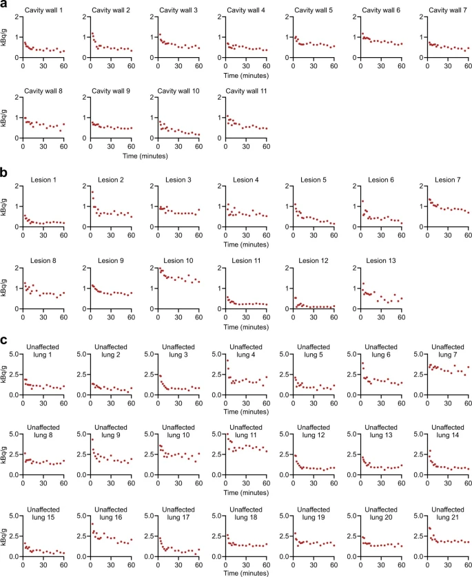
In this recent publication Ordonez et al. provided estimates on rifampin dosing, a first-line TB drug, required to achieve faster cure in 4 months. Newly identified patients were enrolled in a first-in-human study using dynamic [11C]rifampin positron emission tomography (PET) and computed tomography (CT). The findings from human studies were supported with small animal imaging using Mediso nanoScan PET/CT (extended data in publication Fig. 6).
An interesting finding was that [11C]rifampin exposures in human pulmonary TB lesions were low, were spatially compartmentalized and demonstrated between-patient and within-patient variability. [11C]rifampin (tissue-to-plasma) AUC ratios demonstrate limited [11C]rifampin exposure in lesions, with the lowest exposure noted in cavity walls, which paradoxically also have the highest bacterial burden. The pharmacokinetic studies conducted on a rabbit model also demonstrated limited and spatially compartmentalized [11C]rifampin exposures in TB lesions, with the lowest exposures in cavity walls.
The authors hypothesize that tissue necrosis and presumably the fibrotic extracellular matrix surrounding cavitary tissues limited the ability for passive diffusion and rifampin penetration into these lesions, thus minimizing the spread.
Commercially available BSL-3 compliant imaging chamber from Mediso
Mediso has developed in collaboration with Dr. Sanjay K. Jain a commercially available BSL-3 compliant imaging chamber for MultiCell™ imaging system.
The advance of using this chamber is that the PET/CT scanner doesn’t have to be in BSL-3 laboratory, so the animal imaging and the scanner maintenance become much easier. Moreover, the scanner in a such setup can be used for non BSL-3 required animal studies also.
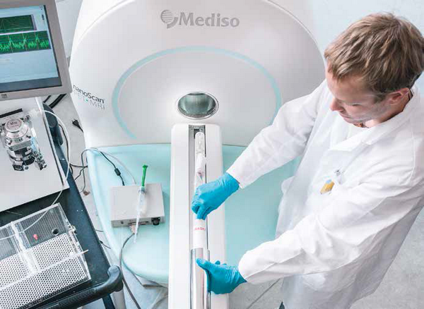
As a workflow, the animals are anesthetized in a BSL-3 environment and placed into the imaging chamber and then sealed. The outer surface of the chamber is decontaminated before caring to BSL-2 environment.
The chamber can be attached by one-click to the scanner’s docking stage without any further tube connection. The anesthesia gas flows in and out through a 0.3 um pore filter. The heating air is circulating only in the chamber’s wall, so there is no transit between the heating air compartment and the chamber space. For kinetic studies the animal is cannulated in BSL-3 and the tubing is led out through a sealed opening (see Figure 1.)
This practical solution provides a tenable and flexible mechanism for routine imaging and for implementing the more severe constraints necessitated by BSL-3 imaging procedures in a simple and expeditious manner.

Figure 1. MultiCell™ BSL-3 compliant imaging chamber

 Coast to Coast
We were thrilled this year to announce that Mediso USA reached a major milestone with the establishment of its tenth preclinical nanoScan imaging system in North America. We are looking back this holiday season with so much appreciation for all of you in making this possible.
 Teaming Up
It was a great honor to have our first site in North America designated as a Center of Excellence for Preclinical Imaging. Many thanks to the Center for Quantitative Cancer Imaging team at the Huntsman Cancer Institute (HCI), part of the University of Utah Health Care system in Salt Lake City. We look forward to continuing our partnership into the New Year.
State of the Art
With its nanoScan PET/MRI(3T) installations dotting the globe, Mediso accepts only the best in imaging performance. As such, the nanoScan PET/MRI(3T) system features a 3T translational MR field strength combined with exceptional PET performance in a compact cryogen-free and low fringe field design, guarantying low running costs and an easy-to-use workflow.
Up and Coming
Our team is also looking forward to a major advance on our horizon. We are proud to say that 2016 will feature our first MultiScan LFER 150 PET/CT installation in the U.S. The large bore in-vivo imaging system is tuned for translational research, capable of whole-body NHP imaging. Time to plan those F220 replacements!
Starting from April 29 the National Institutes of Health (NIH) is now accepting applications for the Shared Instrumentation Grant (SIG) Program and the High End Instrument Grant (HEI) Program.
The submission deadline is May 29, 2015.
The objective of these programs is to make available to institutions expensive, commercially available research systems that cost at least $50,000 (SIG Program) or at least $600,000 (HEI Program). The maximum award is $600,000 for the SIG program and $2,000,000 for the HEI Program.
The instruments can only be justified on a shared-use basis and that are needed for NIH-supported projects in basic, translational or clinical areas of biomedical/behavioral research (description from nih.gov). The SIG Program provides funds to purchase or upgrade a single item of expensive, specialized, commercially available instrument or an integrated instrumentation system to be used for research purposes only. To promote cost effectiveness, to encourage optimal sharing among individual investigators, research groups and departments, and to foster a collaborative multidisciplinary environment, the instrument should be integrated in a centralized core facility, whenever possible.
We, Mediso USA provide support to submit a successful instrumentation grant and we are committed to supporting you throughout the grant process. Please contact us for more details.
External Links
- Shared Instrumentation Grant (SIG) Program (S10): http://grants.nih.gov/grants/guide/pa-files/PAR-15-088.html
- High-End Instrumentation (HEI) Grant Program (S10): http://grants.nih.gov/grants/guide/pa-files/PAR-15-118.html
VivoQuant™ platform for image viewing and analysis 1.23 was released last week by inviCRO.
The VivoQuant™ application is bundled with our nanoScan small animal in vivo imaging systems and software updates are provided for customers with Mediso USA service contracts. As an Imaging Service contract holder, our users not only have access to service and support, but receive the additional capabilities such us upgrading their InVivoScope or VivoQuant software to the new releases.
VivoQuant 1.23 introduces new and improved features, including:
- New Whole Body Atlas Segmentation Tool,
- Deformable Registration Tool,
- Modeling Operator with GLM and Tracer-kinetic Based Modeling,
- Expanded VivoScript Capabilities with New Example Scripts,
- Upgraded 3D ROI Tool with Enhanced Functionality,
- Improved 3D Image Rendering,
- Integration of the Registration / Reorientation Tool with the 3D ROI Tool,
- Improved MR/CT/PET/SPECT Atlas-based Brain Segmentation Plugin - (Sub-region Volume, Signal, fMRI).
To upgrade your VivoQuant version, please go to Tools|Update Check in the software menu or download directly from the VivoQuant website.
There is a nanoScan SPECT/CT in Boston - installed after Thanksgiving - stay tuned for more information.