


Innovation Delivered
 This article was published in discovered, The HZDR Research Magazine (Issue 02.2013, December 2013/January 2014, ISSN: 2194-5713; PDF 2.2MB)
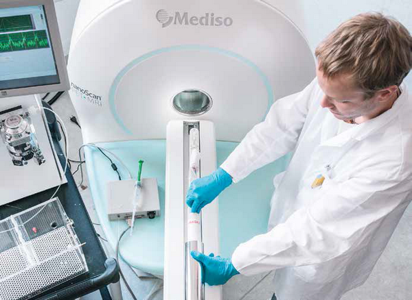
Six white CD-1 mice are scurrying through the litter in their cage, climbing the metal bars, nibbling away at the pellets they are being fed, and snuggling with each other. What they don't yet know is they're about to participate in a pivotal study. One that will save lives - those of mice and, one day, of men. As part of his dissertation, Mathias Kranz, Ph.D. student at the HZDR Research Site Leipzig, is currently investigating the degree of radioactivity that builds up within the bodies of mice whenever radioactive probes - called radiotracers - are used, and identifying in which organs specifically it accumulates. Eventually, these data will be extrapolated to the human magnitude. Radiotracers are chemical compounds that include a radioactive element of some sort, which can help scientists observe metabolic processes in living organisms.
Specifically, in the case of the Leipzig project, we're talking about the two radiotracers [18F]fluspidine and [18F]flubatine - both of them molecules containing the radionuclide 18F (fluorine). They're supposed to ultimately find their way into the diagnostics of cancers and neurodegenerative diseases like Alzheimer's. Key is their ability to imitate properties of various endogenous structures.
Before a radioactive probe is ready for use in the hospital setting, its efficacy and safety must first be documented in living organisms.
Once injected into the human body, they bind with high affinity to certain targets - in the case of the "PET sugar" [18F]FDG, which is also used at the Leipzig site, highly metabolically active tissues like tumors. The emitted radiation from the radioactive molecules can be captured and subsequently analyzed using positron emission tomography (PET). However, before a radioactive tracer can be introduced into the hospital setting, its efficacy and safety to the living organism must first be confirmed. This is a prerequisite imposed by the German Federal Office for Radiation Protection (BfS) and the Federal Institute for Drugs and Medical Devices (BfarM). This multistep procedure starts with work on mice and occasionally pigs and ultimately leads to research conducted on healthy human subjects. Here, the HZDR scientists are receiving support from their colleagues at the Clinic for Nuclear Medicine at Leipzig University Hospital.
Leipzig as reference site
As of spring 2013, when operations by experienced colleagues at the HZDR main site Dresden first commenced, Germany's first-ever commercial full-body PET/MRI for small animals opened in Leipzig - one of only a few worldwide. The HZDR is the reference site for Hungarian manufacturer Mediso (Budapest) - which brings with it a number of obvious benefits: "There are still a handful of delayed-onset childhood illnesses but whenever we do report any problem, help typically arrives within a matter of hours," Mathias Kranz explains. The 27-year-old fellow, who holds a master's in engineering, studied biomedical technology at Ilmenau University of Technology, and has been working at the HZDR Institute of Radiopharmaceutical Cancer Research for about a year now. He is thrilled with the new device: "Not only does it allow us to obtain information about metabolic processes that are happening inside the body, it also yields high-resolution three-dimensional images that document the exact location and distribution of soft tissues." especially when it comes to brain imaging, MR devices yield far better results than conventional PET and computer tomography (CT) combinations.
The mice remain safe
"Without these methods, we would need to dissect the animal subjects, remove individual organs, and then measure them in order to determine the degree of radioactivity that has accumulated in the body following injection of the radiotracer. What's interesting is not only the current dose rate but also how it changes over the course of minutes and hours, which helps determine the organ dose. Thanks to PET/MRI, we're able to conduct even long-term studies using the same exact mouse," Mathias Kranz explains. In the case of other methods, one laboratory animal has to be sacrificed each time a single measurement is obtained.
During examination, the mice are lying on a heated animal bed, their breathing monitored with the help of a pressure sensor. The radioactively labeled substance is injected into the tail vein. The mice are fully anesthetized and won't remember anything afterwards. On a screen, Mathias Kranz is now examining a black and grey image showing the inside of the mouse's body. Red, yellow, and blue spots are lighting up in certain body regions. "Red means these are sites where there is a high degree of radioactivity, in other words that a lot of our substance was deposited in these places," the young scientist explains. At first glance, the liver, kidneys, and bladder are easily recognized - organs, which are actively involved in the substance's elimination from the body.
After the experiments are done, Mathias Kranz calculates the expected effective human dose. This serves as a risk-assessment at the time of introducing the probes into the clinical setting. Based on their results, the researchers have filed for approval of a study with the BfS for use of their newly developed radiotracers (+)-[18F]flubatine and (S)-(-)-[18F]fluspidine in humans. The scientists are working closely with their colleagues at Leipzig University Hospital, Department of Nuclear Medicine, on these projects. The projected start date is early 2014.

Hyperpolarized 13C imaging approach increases the MR signal more than 20,000 times for studying real-time metabolism of disease. Metabolic MRI with hyperpolarized agents shows promise by helping support the differentiation of benign and malignant lesions, separating aggressive from slow-growth tumors and facilitating non-invasive treatments.
The Need for Speed
Molecular Imaging describes techniques that directly or indirectly visualize, characterize, and measure the distribution of molecular or cellular processes at the molecular and cellular levels in humans and other living systems.
The most suitable modalities for small-animal in vivo imaging applications are based on nuclear medicine techniques (essentially, positron emission tomography [PET] and single photon emission computed tomography [SPECT]), optical imaging (OI), computed tomography (CT), magnetic resonance imaging (MRI), magnetic resonance spectroscopy imaging (MRSI), and ultrasound.
Conventional magnetic resonance imaging (MRI) relies on magnetic resonance (MR) signal from proton nuclei of water within the body. The MR signal is encoded with magnetic field gradients for 2D and 3D imaging with no fundamental barriers to spatial resolution as long as sufficient MR signal is available.MRI provides excellent contrast and spatial resolution without radiation exposure - however one limitation of MRI in particular is low sensitivity, especially when compared to PET or SPECT.
Hyperpolarization
Hyperpolarization may address this problem by polarizing spins of a nucleus by several orders of magnitude that seen at thermodynamic equilibrium. However this technique practically doesn't work in water, because spins return back to their equilibrium state, i.e. very low polarization, within seconds. 3He, 13C, 15N, 129Xe and other nuclear spins can be hyperpolarized to the order of near unity resulting in signal enhancement by 4-6 orders of magnitude. Moreover, the decay of their hyperpolarized spin state can be as long as several hours - making useful chemical compounds as hyperpolarized contrast agents. These agents are prepared by physical and/or chemical manipulations followed by administration of these contrast agents in living organisms and their MRI or MRSI imaging.
Hyperpolarized (HP) 129Xe and 3He have been achieved by optical pumping, with potential for low-radiation imaging of the lungs. For nuclei found in endogenous molecules (in particular carbon and nitrogen), the dynamic nuclear polarization (DNP) technique has emerged as a way to polarize small-molecule metabolites. Briefly, 13C-labeled molecules, doped with small quantities of a stable radical, are cooled to approximately 1 K in a magnetic field; microwave irradiation transfers polarization from the fully polarized electron spins on the radical to the 13C nuclei. The sample is then rapidly dissolved using a hot pressurized solution, which can be injected into an animal (or human) in a separate imaging magnet.
Opening the fourth dimension by Chemical Shift Imaging
This approach increases the MR signal more than 20,000 times, thus increasing the biological sensitivity of hyperpolarized MR imaging. Hyperpolarized contrast agents are similar to radioactive tracers in that their signal- generating capability decays exponentially with time - similar to SPECT and PET tracers. The dramatic signal enhancements obtained allow not only the detection of the introduced metabolic agent, but also its metabolic products in real-time. This enabled by magnetic resonance spectroscopic imaging (MRSI) offering the fourth dimension of chemical shift reporting on composition of tissue, i.e. imaging of protons of metabolites in tumors, cardiac tissue and brain, in addition to three spatial dimensions. Its biggest application so far has been in imaging the glucose consumption in tumors — glucose and lactate for the localization of benign and malignant prostate cancer. this concept has a lot of potential for other kinds of metabolic applications, too, most notably diabetes imaging.
Despite signal boost by several orders of magnitude, hyperpolarized MRI relies on signal from relatively dilute spins of administered hyperpolarized contrast agents. For example, hyperpolarized 13C-lactate concentration in vivo is on the order of a few mM, which is several orders of magnitude lower than proton concentration of tissue water. As a result, SPECT and PET are inherently significantly more sensitive (by orders of magnitude) imaging modalities when accounting for contrast agent quantity. When comparing hyperpolarized MRI to PET imaging, it should also be noted that the vast majority of hyperpolarized contrast agents have significantly shorter lifetime on the order, of 0.5-5 minutes in vivo. This double-edged sword limits the use of hyperpolarized contrast agents from the perspective of metabolic pathways penetration, contrast agent in vivo delivery, pharmaceutical preparation and imaging site distribution. On the other hand, it offers an opportunity to perform a repeat scan within minutes after initial hyperpolarized scan, because there is no background signal from the first initially administered dose.
Bringing it into one system
PET/MR imaging is just a phenomenal tool — it combines two very strong technologies. This field however opens even more new opportunities by potentially combining the power of molecular imaging of hyperpolarized MRI and high sensitivity PET. While the main advantage of hyperpolarized MRI is the large sensitivity boost enabled by increased nuclear spin polarization, this increase is not endowed by the magnetic field of the MRI scanner. As a result, it is possible to perform MRI of hyperpolarized contrast agents in very low magnetic fields. The nanoScan PET/MRI is equipped with a permanent 1T magnet which is seamlessly integrated and automated into the equipment. Our advantage is the inherently low cost maintenance, because the need for a high-field cryogenic magnet is eliminated and also no other site preparation and supportive maintenance, like water cooling is required. The combination of low cost and sub-second scan speed is a clear advantage.
Further readings
The hyperpolarized MRI is and emerging and quickly developing field, however its importance can assessed by the increasing number of published articles and presentations on conferences. Recently a review article was published on 13C hyperpolarized magnetic resonance using dynamic nuclear polarization in Chemical Society Reviews written by Kayvan R. Keshari and David M. Wilson.
Suggested literature
- Keshari, Kayvan R., and David M. Wilson. "Chemistry and Biochemistry of 13C Hyperpolarized Magnetic Resonance Using Dynamic Nuclear Polarization." Chemical Society Reviews 43, no. 5 (February 10, 2014): 1627–59. doi:10.1039/C3CS60124B.
- Gallagher, Ferdia A., Sarah E. Bohndiek, Mikko I. Kettunen, David Y. Lewis, Dmitry Soloviev, and Kevin M. Brindle. "Hyperpolarized 13C MRI and PET: In Vivo Tumor Biochemistry." Journal of Nuclear Medicine 52, no. 9 (September 1, 2011): 1333–36. doi:10.2967/jnumed.110.085258.
- Chekmenev, Eduard Y. MRI "Hyperpolarization and Molecular Imaging" mi Gateway, Newsletter of the SNMMI CMIIT, Vol. 7, Issue 3, 2013-3
The suggested reading list was actually used to prepare this post. This was an introductory post in the realm of HP MRI imaging - hope you enjoyed it.
There is a nanoScan SPECT/CT in Boston - installed after Thanksgiving - stay tuned for more information.
Last month, in October a new review article titled Preclinical Imaging: an Essential Ally in Modern Biosciences on preclinical imaging technologies was published in the Molecular Diagnosis & Therapy journal. The journal provides insights into the latest molecular diagnostic and pharmacogenomic techniques and their use in personalized medicine.
Cunha, Lídia, Ildiko Horvath, Sara Ferreira, Joana Lemos, Pedro Costa, Domingos Vieira, Dániel S. Veres, et al. 2013. “Preclinical Imaging: An Essential Ally in Modern Biosciences.” Molecular Diagnosis & Therapy: 1–21. doi:10.1007/s40291-013-0062-3.
The find out that actually what is small-animal or preclinical imaging, P. Zanzonico from MSKCC has provided a good definition, stating that 'it constitutes a way of assessing biological structures and function in vivo by noninvasive means, allowing the collection of quantitative information, both in health and disease states' [1].
The main role of preclinical imaging is to deliver translational answers for serious health-related problems of the growing and aging world population. Small animal models have to represent a bridge between discoveries at the molecular level and clinical implementation in diagnostics or therapeutics. Small animal imaging is being used in a wide variety of lines of research, especially in infection, inflammation, oncology, cardiology, and neurosciences.
The article summarizes the general properties of diagnostic imaging modalities and reviews them one-by-one including Positron emission tomography (PET), Single photon emission computed tomography (SPECT), Optical imaging (OI), Computed tomography (CT), Magnetic resonance imaging (MRI) and Ultrasound (US) and their related instrumentation of these modalities in small animal imaging. A separate and well detailed section is dedicated to the comparison of micro-SPECT and micro-PET. The general parameters are summarized in a large table listing imaging characteristics (spatial resolution, sensitivity, penetration depth, temporal resolution), related costs, probe types, major advantages, disadvantages and their application areas.
There are inherent limitations to each imaging modality - this has brought commercial multi-modality systems 10+ years ago to the market. Multimodal combination has enabled some of the most important limitations of each imaging modality to be overcome when used alone. The considerations are explained in the tenth sections of the article.
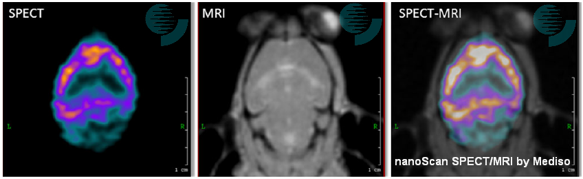
It's an honor to see multi-modality images of PET/MRI and SPECT/MRI acquired by our nanoScan imagers in the article.
A SPECT/MRI application was selected as the image of this blog post. The image shows transverse slices of SPECT and MRI images of a mouse brain. SPECT was acquired using a specific agent for cortical benzodiazepine receptors (123I-NNC13-82431). The lack of anatomical information of SPECT acquisition is complemented with the information provided by MRI, in which the eyes, the olfactory bulbs and the first and second ventricles are shown. The multimodality SPECT/MRI image provides information about functional benzodiazepine receptors from SPECT allied to good soft tissue contrast from the MRI.
Abstract of the Article
Translational research is changing the practice of modern medicine and the way in which health problems are approached and solved. The use of small-animal models in basic and preclinical sciences is a major keystone for these kinds of research and development strategies, representing a bridge between discoveries at the molecular level and clinical implementation in diagnostics and/or therapeutics. The development of high-resolution in vivo imaging technologies provides a unique opportunity for studying disease in real time, in a quantitative way, at the molecular level, along with the ability to repeatedly and non-invasively monitor disease progression or response to treatment. The greatest advantages of preclinical imaging techniques include the reduction of biological variability and the opportunity to acquire, in continuity, an impressive amount of unique information (without interfering with the biological process under study) in distinct forms, repeated or modulated as needed, along with the substantial reduction in the number of animals required for a particular study, fully complying with 3R (Replacement, Reduction and Refinement) policies. The most suitable modalities for small-animal in vivo imaging applications are based on nuclear medicine techniques (essentially, positron emission tomography [PET] and single photon emission computed tomography [SPECT]), optical imaging (OI), computed tomography (CT), magnetic resonance imaging (MRI), magnetic resonance spectroscopy imaging (MRSI), and ultrasound. Each modality has intrinsic advantages and limitations. More recently, aiming to overcome the inherent limitations of each imaging modality, multimodality devices designed to provide complementary information upon the pathophysiological process under study have gained popularity. The combination of high-resolution modalities, like micro-CT or micro-MRI, with highly sensitive techniques providing functional information, such as micro-PET or micro-SPECT, will continue to broaden the horizons of research in such key areas as infection, oncology, cardiology, and neurology, contributing not only to the understanding of the underlying mechanisms of disease, but also providing efficient and unique tools for evaluating new chemical entities and candidate drugs. The added value of small-animal imaging techniques has driven their increasing use by pharmaceutical companies, contract research organizations, and research institutions.
[1] Zanzonico P. Noninvasive imaging for supporting basic research. In: Kiessling F, Pichler BJ, editors. Small animal imaging—basics and practical guide. Heidelberg: Springer; 2011. p. 3–16. (Springer; Google Books)

A New Spin on the Story of AnyScan
as illustrated by Gergo P.
Lego bricks give the limitless possibilities represented by an unassembled pile. You can build just about anything out of LEGOs these day if you've got the patience and enough bricks.
And this happened with the AnyScan product family of Mediso as Gergo, one of our magnificent physicists (a clear magician in GATE Monte Carlo simulations) created his own slightly greater-than-minifig-scale of the Mediso AnyScan PET-SPECT-CT clinical tomographic scanner.
The album can be reached on Flickr.
And the fun part is that the AnyScan S, single-head and dual-head large field-of-view general purpose SPECT camera just received the FDA 510(k) clearance, bringing this great medical device to the US market.
The AnyScan® S is a proven 4th generation system with installations around the world, and offers a unique solution in molecular imaging with an ergonomic open design gantry, variable angle detector positions, small footprint, robust mechanical design with improved safety factor, dual infrared line auto body contouring, total body localizer mode, table design to support patients up to 500 lbs., and pre-programmed robotic gantry motions with full automatic motion positioning and calibration. In addition, the flexible modular system architecture provides a pathway to offer variety of modalities within the AnyScan® family.
Back in May 2013, I gave a talk titled "The Motivations and Systems for High Content In-vivo Tomographic Imaging in
Drug Discovery" at the 6th Imaging in Drug Discovery & Development Conference in Boston. Mediso USA was the Silver Sponsor for the event.
According to GTC this is "the only imaging conference that brings together high-level/influential leaders with decision-making authority from the pharmaceutical industry, academia, and government to share their knowledge and expertise in drug discovery and development". Needles to say, the sessions were indeed interesting, with an interesting mix from academia, government, pharma and imaging companies.
Session topics included:
- Advantages and Challenges of Available Imaging Modalities
- Translational Imaging Applications: Preclinical to Clinical
- Imaging Applications Across Multiple Therapeutic Areas
- Molecular Imaging and Diagnostic Approaches and Capabilities
- High Content Imaging, Quantitative Imaging and Modeling Capabilities
The November issue of the was published today in the Genetic Engineering & Biotechnology News, with the Feature Article: Raising the Bar in Preclinical Imaging written by MaryAnn Labant. The article is based on presentations given at the May GTC Imaging in Drug Discovery and Development Conference.
Please find below our related section from the second page of the online article.
Integrated Imaging Systems
Preclinical PET scanners with an integrated microCT have substantially improved the anatomical registration of PET predominately to the skeleton, yet little progress has been made in soft tissue contrast, even with the use of a CT contrast agent.
 Integrated PET/MRI or SPECT/MRI systems offer many benefits. MRI uses no radiation, offers better soft tissue contrast, and provides molecular readouts. To date, preclinical PET imaging combined with MRI has been performed using two independent systems and a bespoke co-registration algorithm to fuse the images.
Mediso recently commercialized the first serially produced, fully integrated, automated PET/MRI system, the nanoScan PET/MRI, and a fully integrated, automated SPECT/MRI system, the nanoScan SPECT/MRI. Single systems enable use of the same imaging technology, imaging protocol, and biomarkers for small to large subjects.
According to Illes J. Muller, managing partner, preclinical PET/MRI and SPECT/MRI allow combination of radionuclide biomarkers with an MRI contrast agent on a routine basis, an attractive prospect for evaluating new drugs for oncology, neurology, and cardiovascular disease. Now, physiological/metabolic readouts can be combined with high-resolution, soft-tissue contrast as well as dynamic functional perfusion imaging.
 In addition, SPECT provides the ability to perform multi-isotope imaging, probing two or more molecular pathways simultaneously by detecting isotopes with different emission energies, and has no physical limits in resolution. SPECT/MRI technology is less expensive. The labeling is easier, and no on-site cyclotron is required.
A potential major application for multimodal emission tomography combined with MRI is quantitative 3D imaging of tumor heterogeneity. To assess the spatial distribution of a given PET or SPECT biomarker within a tumor requires ultra-high resolution and high sensitivity and corrections for tumor perfusion. MRI is able to differentiate between healthy and dead tumor tissue for tumor response evaluation.
Note: This was the related section from the article, with links added to the relevant pages of Mediso USA website.
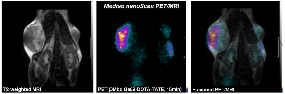
Blog Image
The blog image shows a Mouse Tumor Heterogeneity Study performed with nanoScan PET/MRI. The mouse was injected with 3 MBq Ga68-DOTA-TATE and imaged for 15 minutes at 60-75 min post injection.
Please join us at the Fairmont Copley Plaza in Boston for the second annual Advanced Molecular Imaging and its Clinical Translation course, starting this Sunday, Oct. 27 and continuing through Wednesday, Oct. 30.
Mediso USA is proud to be a sponsor of this important course.
The course will provide a comprehensive overview in the physics, chemistry, engineering, and physiology that are the foundation of molecular imaging. It will cover SPECT, PET, CT, MRI, optical imaging, ultrasound, multi-modality imaging, contrast agent chemistry, radiotracer development, preclinical imaging, issues surrounding clinical translation, and other aspects of molecular imaging.
Course Directors are:
John V. Frangioni, M.D., Ph.D.: Professor of Medicine and Professor of Radiology; Director, Center for Molecular Imaging, Beth Israel Deconess Medical Center, Boston, MA.
Ralph Weissleder, M.D., Ph.D.: Professor of Radiology and Systems Biology; Massachusetts General Hospital, Boston, MA.
Dr. Frangioni's lab includes both a NanoSPECT/CT and a NanoPET/CT small animal imaging system, built by Mediso.
We hope that you are able to attend and that you will visit us at the exhibit hall.
The SNMMI is now accepting abstract submissions for the upcoming SNMMI 2014 Annual Meeting.The congress will be held between June 7-11, 2014 in St. Louis, Missouri.
The abstract submission deadline is Friday, January 3, 2014 (except Technologist Student Submissions). Practically it means that either you will have to write before New Years Eve or you will be willing to work on it right after New Years Eve.
The abstracts should be submitted through the meeting's Submission Website. Detailed information about the application process can be downloaded in PDFs from the SNM website.
----
Update: Mediso USA will exhibit at booth #1421 at the SNMMI 2014 Annual Congress. Come and see us!
The European Association of Nuclear Medicine (EANM) is the umbrella organisation of nuclear medicine in Europe and represents the sector towards the European Institutions. The EANM 2013 Annual Meeting will be held in Lyon, France this year in October. Visit the related EANM 2013 Event Page on the Mediso USA website for more details.
Update: Invitation for the User Meeting (October 21, 1:00-3:00 PM).
Before reading all details on our exhibit and programs, watch the video below about Lyon, a UNESCO World Heritage Site.

I'm excited to announce that we've started our blog. Just two days before the opening of WMIC 2013 in Savannah, the first post lists the selected presentations citing the Mediso systems at this conference.
Download the listing in PDF format.
Thursday
In Vivo Imaging of Microglia Cells Activated by LPS-Induced Systemic Inflammation in Mouse
Domokos Mathe1, Ildiko Futo2, Daniel Veres2, Ildiko Horvath2, Mariann Semjeni1, Noemi Kovacs1, Miklós Tóth3, Ralf K. Bergmann4, Krisztian Szigeti1
1. CROmed Ltd, Budapest, Hungary. 2. Biophysics and Radiation Biology, Semmelweis University , Budapest, Hungary. 3. Clinical Neuroscience, Karolinska Institute, Stockholm, Sweden. 4. Radiopharmacy Radiopharmaceutical Biology, Helmholtz-Zentrum, Dresden-Rossendorf, Germany.
| P218. Thursday, Sept 19, 15:15-16:45 Poster Session 2 (Exhibit Hall B) |
nanoScan PET/MRI Silver Upgrade of NanoSPECT/CT |
89Zr-Oxine Complex: a Long-Lived Radiolabel for Cell Tracking Using PET
Levente K. Meszaros1, Putthiporn Charoenphun1, Krisanat Chuamsaamarkkee1, James R. Ballinger1, 2, Greg Mullen1, Trevor J. Ferris3, Michael J. Went3, Phil Blower1
1. Department of Imaging Chemistry and Biology, King's College London, London, United Kingdom. 2. Department of Nuclear Medicine, Guy’s and St Thomas NHS Foundation Trust, London, United Kingdom. 3. School of Physical Sciences, University of Kent, Canterbury, Canterbury, United Kingdom.
| LB024. Thursday, Sept 19, 15:15-16:45 Late Br.Abst.Poster S (Exhibit Hall B) |
nanoScan PET/CT |
PSMA Specific Diabody For SPECT Imaging of Prostate Cancer
Florian Kampmeier1, Jennifer D. Williams1, John Maher2, 3, 4, Greg Mullen1, Phil Blower1
1. School of Medicine, Division of Imaging Sciences & Biomedical Engineering, King's College London, London, London, United Kingdom. 2. King’s Health Partners Integrated Cancer Center, Department of Research Oncology, King's College London, London, London, United Kingdom. 3. Department of Immunology, Barnet and Chase Farm NHS Trust, London, London, United Kingdom. 4. Department of Clinical Immunology and Allergy, King’s College Hospital NHS Foundation Trust, London, London, United Kingdom.
|
LBAP105. Thursday, Sept 19, 15:15-16:45 |
nanoScan PET/CT |
Spatio-Temporal Quantification of Sodium Iodide Symporter (NIS) Radiotracers using pre-clinical SPECT/CT and PET/CT: A Study in Healthy Scid/Beige Mouse
Krisanat Chuamsaamarkkee1, Seckou Diocou1, Greg Mullen1, Lefteris Livieratos1, 2, Phil Blower1
1. Imaging Sciences and Biomedical Engineering, King's College London, London, United Kingdom. 2. Nuclear Medicine, Guy’s & St Thomas’ Hospitals NHS Foundation Trust, London, United Kingdom.
|
LBAP106. Thursday, Sept 19, 15:15-16:45 |
Silver Upgrade of NanoSPECT/CT |
Friday
Study of Acetyl-L-Carnitine Effects on Glucose Metabolism in Mouse Brain Using PET/MRI Imaging
Lidia S. Cunha3, 4, Domokos Mathe1, Ildiko Horvath2, Daniel Veres2, Krisztián Szigeti2, Luis F. Metello3, Teresa Summavieille4
1. CROmed Ltd, Budapest, Hungary. 2. Biophysics and Radiation Biology, Semmelweis University, Budapest, Hungary. 3. Nuclear Medicine Department, High Institute for Allied Health Technologies of Porto - Polytechnic Institute of Porto, Porto, Portugal. 4. Neuroprotection Lab, IBMC, University of Porto, Porto, Portugal.
|
P341. Friday, Sept 20, 15:15-16:45 |
nanoScan PET/MRI |
In vivo basic tracer pharmacokinetic analysis for transgenic mouse models of Alzheimer's disease
Krisztián Szigeti1, Gellért- Szabolcs Kovács1, István Varsányi1, Ferenc Budán2, Ildiko Horvath1, Albert D. Windhorst3, Domokos Mathe2
1. Biophysics and Radiation Biology, Semmelweis University, Budapest, Hungary. 2. CROmed Ltd, Budapest, Hungary. 3. Radiology, Nuclear Medicine and PET Research, Vrije University, Amsterdam, Netherlands.
|
P349. Friday, Sept 20, 15:15-16:45 |
nanoScan PET/MRI |
Saturday
Dual modality optical and radionuclide reporter gene imaging of heterogeneous chemotherapy response in different microenvironments
Gilbert O. Fruhwirth1, 3, Seckou Diocou1, Phil Blower1, 3, Tony Ng2, 3, Greg Mullen1
1. Department for Imaging Chemistry and Biology, King's College London, London, United Kingdom; 2. Dimbleby Department of Cancer Research, King's College London, London, United Kingdom; 3. Comprehensive Cancer Imaging Centre KCL & UCL, King's College London, London, United Kingdom.
|
SS119. Saturday, Sept 21, 11:30-11:45 |
Silver Upgrade of NanoSPECT/CT |
PEGylated Core/shell Doped Fe3O4@NaYF4 Nanoparticles: Multimodality Molecular Imaging Contrast for MRI, PET/SPECT and Optical Imaging
Xianjin Cui1, Phil Blower1, 2, Mark Green1, 3
1. Imaging Science, King's College London, London, United Kingdom. 2. Chemistry, Kings College London, London, United Kingdom. 3. Physics, Kings College London, London, United Kingdom.
|
P450. Saturday, Sept 21, 14:45-16:15 |
nanoScan PET/CT |
Lessons learnt from reporter gene imaging of regulatory T cell therapy in transplantation: a non-invasive whole body nuclear imaging study.
Ehsan Sharif-Paghaleh1, 2, John Leech1, Robert Lechler1, Lesley A. Smyth1, Giovanna Lombardi1, Greg Mullen1, 2
1. MRC Centre for Transplantation, KCL, London, United Kingdom. 2. Imaging Sciences, KCL, London, United Kingdom.
|
P525. Saturday, Sept 21, 14:45-16:15 |
Silver Upgrade of NanoSPECT/CT |
PET/MRI/SPECT/CT in vivo longitudinal imaging of Earthworm (Lumbricus terrestris L.), as a novel means of environmental monitoring
Ferenc Budán2, Noemi Kovacs2, Ildiko Horvath1, Daniel Veres1, Péter Engelmann3, Peter Nemeth3, Krisztián Szigeti1, Domokos Mathe2
1. Biophysics and Radiation Biology, Semmelweis University, Budapest, Hungary. 2. CROmed Ltd, Budapest, Hungary. 3. Department of Immunology and Biotechnology, University of Pécs, Pécs, Hungary.
|
P535. Saturday, Sept 21, 14:45-16:15 |
nanoScan PET/MRI |
Minimum Detectable Activity of a Preclinical PET/MR System
Kalman L. Nagy1, 2, Judit Lantos1, Péter Major1, Gergely Patay1, Christer Halldin2, Balázs Gulyás2
1. Mediso ltd, Budapest, Hungary. 2. Dept. Clinical Neuroscience, Karolinska Institutet, Stockholm, Sweden.
|
P603. Saturday, Sept 21, 14:45-16:15 |
nanoScan PET/MRI |
We're looking forward to meeting with you, please come to our booth to get this listing in printed format. If you cannot make it this year, please feel free to contact us for copies of all of these posters (after they have been presented).
By accepting you will be accessing a service provided by a third-party external to https://medisousa.com/dev/